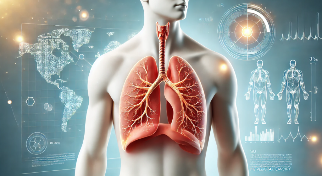
Pneumothorax is a pathological condition in which air enters the pleural cavity, leading to lung compression and impaired function. This condition can be life-threatening and requires immediate medical attention.
1. Mechanism of Pneumothorax Development
The pleural cavity is the space between the lung and the chest wall, normally filled with a small amount of fluid that facilitates smooth lung movement. In pneumothorax, air enters this space, increasing pressure and preventing the lung from expanding during inhalation.
2. Types of Pneumothorax
- Spontaneous Pneumothorax: Occurs without apparent cause, often in individuals with underlying lung conditions like bullous emphysema.
- Traumatic Pneumothorax: Develops as a result of chest injuries, such as rib fractures or penetrating wounds.
- Iatrogenic Pneumothorax: May occur due to medical procedures, such as catheter placement.
- Tension Pneumothorax: The most dangerous form, where air continues to accumulate, increasing pressure on chest organs.
3. Main Symptoms
- Shortness of Breath: Impaired breathing due to lung compression.
- Chest Pain: Typically sharp and worsens with inhalation.
- Cyanosis: Bluish lips and skin due to oxygen deficiency.
- Tachycardia: Rapid heartbeat in response to oxygen deprivation.
- Chest Asymmetry: The affected side may show reduced movement during breathing.
4. Diagnosis

- Chest X-ray: Detects air in the pleural cavity.
- CT Scan: Used for detailed diagnosis and lung tissue assessment.
- Pulse Oximetry: Measures oxygen levels in the blood.
5. Treatment Methods

- Small Spontaneous Pneumothorax: May be treated conservatively with observation and oxygen therapy.
- Pleural Drainage: Involves inserting a tube to remove air.
- Surgery: Required for recurrent pneumothorax or traumatic injuries.
- Emergency Care: In tension pneumothorax, immediate chest puncture is performed to relieve pressure.
6. Prevention
- Avoid smoking, which damages lung tissue.
- Timely treatment of lung diseases.
- Use protective gear during extreme sports.
Conclusion
Pneumothorax is a serious condition that requires prompt identification and treatment. Paying close attention to your health and seeking timely medical care can help prevent complications and maintain well-being.
Pneumothorax: Understanding the Condition and Its Management
Pneumothorax, commonly referred to as a collapsed lung, is a medical condition that occurs when air enters the space between the lung and the chest wall, known as the pleural space. This air buildup creates pressure on the lung, causing it to partially or completely collapse. Pneumothorax can range from a minor issue that resolves on its own to a life-threatening emergency requiring immediate medical intervention. Understanding the causes, symptoms, and treatments of pneumothorax is crucial for prompt diagnosis and effective management.
There are several types of pneumothorax, each with distinct causes and characteristics. Spontaneous pneumothorax occurs without an apparent injury or trauma and is further classified into primary and secondary types. Primary spontaneous pneumothorax typically affects individuals without underlying lung disease, often young, tall men, and smokers. Secondary spontaneous pneumothorax occurs in people with pre-existing lung conditions such as chronic obstructive pulmonary disease (COPD), asthma, cystic fibrosis, or tuberculosis.
Traumatic pneumothorax results from an injury to the chest, such as a fractured rib, gunshot, or stab wound. Medical procedures like lung biopsies or the insertion of a central venous catheter can also cause traumatic pneumothorax. Tension pneumothorax, a severe and potentially life-threatening form, occurs when air continues to enter the pleural space without an escape route, leading to increased pressure on the lung, heart, and major blood vessels. This can cause a rapid decline in respiratory and cardiovascular function and requires immediate intervention.
The symptoms of pneumothorax vary depending on its size and severity. Common signs include sudden chest pain, which may be sharp or stabbing, and shortness of breath. Other symptoms can include rapid breathing, a rapid heart rate, fatigue, and a feeling of tightness in the chest. In severe cases, cyanosis (bluish discoloration of the skin), low blood pressure, and confusion may occur, particularly with tension pneumothorax. Symptoms often develop quickly, making it essential to seek medical attention promptly.
Diagnosing pneumothorax typically involves a combination of physical examination and imaging tests. During the examination, a doctor may detect diminished or absent breath sounds on the affected side, or hear abnormal sounds during percussion of the chest. A chest X-ray is the most common imaging method used to confirm the diagnosis, showing air in the pleural space and a collapsed lung. In some cases, a CT scan may be required to provide more detailed images, especially in patients with complex symptoms or underlying lung conditions.
Treatment for pneumothorax depends on its type, size, and the patient’s overall health. For small, uncomplicated cases, observation may be sufficient, as the air in the pleural space can be reabsorbed naturally over time. Patients are typically advised to rest and avoid strenuous activities during this period.
For larger or more severe pneumothorax, medical intervention is required. Needle aspiration or the insertion of a chest tube is commonly performed to remove air from the pleural space and allow the lung to re-expand. The chest tube remains in place for a few days to ensure the lung stays inflated while healing occurs. In cases of tension pneumothorax, immediate needle decompression is performed to relieve pressure, followed by chest tube placement.
For recurrent or persistent pneumothorax, surgical intervention may be necessary. A procedure called pleurodesis involves creating a bond between the lung and the chest wall to prevent future air leaks. Another surgical option is video-assisted thoracoscopic surgery (VATS), which can repair lung damage or remove air blisters (blebs) that are prone to rupture.
Recovery from pneumothorax varies depending on its severity and the treatment method. Most patients recover fully, but lifestyle adjustments may be needed to reduce the risk of recurrence. Smoking cessation is strongly recommended, as smoking significantly increases the likelihood of pneumothorax, particularly in those who have already experienced it. Avoiding high-altitude travel, scuba diving, or other activities that involve pressure changes is also advised, as these can exacerbate the condition.
In some cases, pneumothorax can lead to complications, including recurrent episodes, infection, or scarring in the pleural space. People with underlying lung diseases are more susceptible to complications and should work closely with their healthcare providers to monitor and manage their condition.
In conclusion, pneumothorax is a serious medical condition that requires timely diagnosis and appropriate treatment to prevent complications. By understanding its causes, symptoms, and management options, individuals can take proactive steps to minimize risks and seek prompt medical care when needed. Advances in medical technology and treatment approaches have significantly improved outcomes for patients with pneumothorax, offering hope for a full recovery and a return to normal life.


Can you be more specific about the content of your article? After reading it, I still have some doubts. Hope you can help me.